Adherent Electroporation Applications
Cell Culture Transfection
Transfection in Cell-Culture-Plates/Dishes
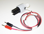
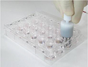
By using the cell-culture-plate Electrode CUY900 series with the NEPA21 electroporator, it is now possible to transfer DNA/RNA directly into cells IN ADHERENCE in a commercially available multi-well plate.
See the cell images by clicking the cell names.
V: Viability, TE: Transfection Efficiency.
| Cells | V | TE | Cells | V | TE | |
| Primary Human Skin Fibroblasts | 100% | 50% | Primary HUVEC | 75% | 65% | |
| Primary Mouse Hippocampal Neurons (Embryonic day 14) (4 DIV) |
60% | 50% | Primary Mouse Hippocampal Neurons (Embryonic day 18) (2 DIV) |
85% | 54% | |
| Mouse Neural Stem Cells | 71% | 50% | Primary Mouse Microglial Cells (1 DIV after 1 week co-culturing astrocyte and microglial cells) |
80% | 73% | |
| Primary Mouse Glial Cells (14 DIV) |
80% | 50% | Primary Mouse Stromal Cells (1-month cultured) |
90% | 50% | |
| Primary Mouse Liver Cells siRNA Knock Down |
Excel. | 89% | ||||
| Primary Rat Cerebral Cortex Neurons (Embryonic day 17) (2 DIV) |
70% | 60% | Primary Rat Hippocampal Neurons (Postnatal day 7) (11 DIV) |
100% | 50% | |
| Primary Rat Granulosa Cells | Excel. | 41% | ||||
| hMSC – Human Mesenchymal Stem Cells | 90% | 70% | SH-SY5Y – Human Neuroblastoma Cells | 90% | 50% | |
| EPC – Human Endothelial Progenitor Cells | HPDE – Human Pancreatic Duct Epithelial Cells | 80% | ||||
| THP-1 – Human Acute Monocytic Leukemia Cells | 90% | 45% | ||||
| C2C12 – Mouse Myotubes | 94% | 60% | 3T3-L1 – Mouse Embryonic Fibroblasts (7 days after differentiation) | 90% | 70% | |
| MEF – Mouse Embryonic Fibroblasts | 80% | 90% | Neuro-2a – Mouse Neuroblastoma Cells | 80% | 90% | |
| C6 – Rat Glioma Cells | 57% | 55% |
Transfection into Primary Neurons
 |
 |
 |
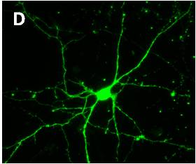 |
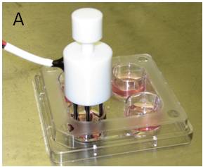
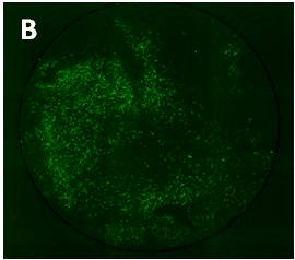
pCAGGS-EGFP plasmid was transferred into primary neurons cultured for 6 days in adherent state.
The neurons were prepared from E15 mouse cerebral cortex.
| A): | 2 steps pulse electroporation using the electrodes (CUY900-13-3-5) for adherent cells |
| B): | EGFP fluorescence image of the neurons 2 days after electroporation |
| C): | High magnification image of Figure B. Many robust EGFP signals suggest high transfection efficiency. |
| D): | High magnification image of Figure C (x40). Neurites are shown clearly. |
| Data provided courtesy of Department of Neurochemistry, National Institute of Neuroscience, Japan | |
We have a lot of data of adherent-cell transfection with high efficiency and high viability. Please feel to free to contact us for the latest data.