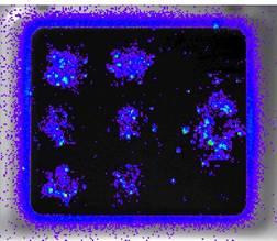
Cells have been plated throughout the window of the unit above and microbubbles together with luciferase-encoding naked plasmid DNA have been added. Areas exhibiting luciferase activity have been treated with ultrasound externally applied to the unit. The data indicate both the non-invasive nature of the ultrasound-based stimulus AND the site-specific nature of gene transfer/expression.
System advantages
• Target cells covered the complete Opticell membrane surface and positions treated with ultrasound are non-destructively visualised by photonic imaging.
• Non-invasive specific spatial targeting of gene transfer to pre-defined sites on the membrane are clearly visible
• Gene expression may be quantified directly using photonic imaging or by excision of the membrane and recovery of the cells
• Gene expression may also be visualised and mapped using fluorescence microscopy if a GFP-based reporter is employed